Just out in today's Science: "Technological challenges and milestones for writing genomes." One of a pair of papers I've been working on with the GP-write consortium, both of which are asking the question: what, exactly, do we need in order to go from engineering millions of base-pairs of DNA in bacteria and yeast to the billions of base-pairs in complex organisms like mammals, plants, and people?
This paper focuses on the DNA-wrangling side of the problem, while its complement (on arXiv and under revision) focuses on the informational and coordination side of the problem. Both need to be addressed, and the complexity---while daunting---is tractable. Take a read-through and see our take on the matter!
Friday, October 18, 2019
Monday, October 14, 2019
Getting plate readers right
If you ever used a plate reader to measure either OD or fluorescence, you'll want to check out the iGEM 2018 interlab preprint on bioRxiv!
We just submitted this manuscript, "Robust Estimation of Bacterial Cell Count from Optical Density," for review on Friday, but we think a lot of folks will want to make use of this information, and so we've gotten a preprint up early as well. The big deal of this study is that we've now got a good calibration process for both optical density (OD) measurement, which is commonly used for estimating cell count in a sample, and fluorescence measurement, which is commonly used as a "debugging probe" for estimating cellular activity. Both of these are usually reported in relative or arbitrary units right now, which causes lots of trouble interpreting what's even going on in your experiment, as well as greatly limiting how results can be shared and applied.
No more: we have protocols that are cheap (less than $0.10/run) and easy (reliably executed by high school students just getting started in a lab), and that this manuscript shows are also both precise and accurate. All you have to do is dilute little cell-sized silica beads and fluorescent dye, plug the measurements into a spreadsheet, and you're good to go.
And here's the most important result from our paper: a nearly perfect match between per-cell fluorescence estimate from plate reader measurements and the ground truth captured from single-cell measurements in flow cytometers.
In fact, this match is even better than we deserve: we know there are factors that should distort the plate reader measurements both up and down, but they're small and appear to be canceling one another out. The only device with a notable difference in measured value is the one that's got very low fluorescence---and even there it's not significant and conforms with our expectation that flow cytometers will be better able to measure extremely faint fluorescence than plate readers.
We just submitted this manuscript, "Robust Estimation of Bacterial Cell Count from Optical Density," for review on Friday, but we think a lot of folks will want to make use of this information, and so we've gotten a preprint up early as well. The big deal of this study is that we've now got a good calibration process for both optical density (OD) measurement, which is commonly used for estimating cell count in a sample, and fluorescence measurement, which is commonly used as a "debugging probe" for estimating cellular activity. Both of these are usually reported in relative or arbitrary units right now, which causes lots of trouble interpreting what's even going on in your experiment, as well as greatly limiting how results can be shared and applied.
No more: we have protocols that are cheap (less than $0.10/run) and easy (reliably executed by high school students just getting started in a lab), and that this manuscript shows are also both precise and accurate. All you have to do is dilute little cell-sized silica beads and fluorescent dye, plug the measurements into a spreadsheet, and you're good to go.
 |
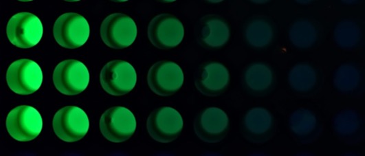
| Serial dilution of fluorescein (from iGEM protocols page) |
And here's the most important result from our paper: a nearly perfect match between per-cell fluorescence estimate from plate reader measurements and the ground truth captured from single-cell measurements in flow cytometers.
 |
| Plate reader (calibrated with microsphere dilution) vs. flow cytometry showing a 1.07-fold mean difference over 6 test devices. |
This is new science, so there's lots of caveats, of course: this has only been validated for E. coli, and probably won't work well for murky cultures with a lot of background or for biofilms or long filamentous strands. Nevertheless, it's a big step forward, since a huge amount of what people use plate readers for is covered by this study already. We'll see what the reviewers think, but I expect this paper is going to have a big impact because it's addressing a problem that so many people are encountering.
The next key challenge, however, is this: can we get somebody manufacturing plate readers to make calibration plates so that people don't have to prepare their reference materials themselves?
Subscribe to:
Comments (Atom)